ACCURACY
Out of 100 cases , 6 more correctly classified
77.6%Pathologists alone
83.9%

Review the tumor regions and cell counts behind CPS and TPS to check how the score was calculated.
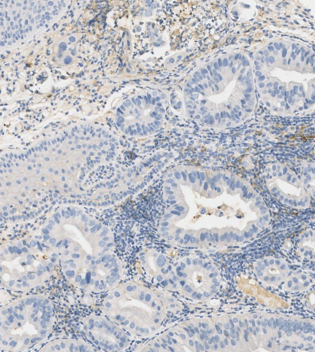
VALIDATED SCORING SCOPE
HOW IT WORKS
Access the slide for quantification directly through your supported image management system.
Review the tumor regions and cell counts behind CPS and TPS to check how the score was calculated.
The assay returns the score and supporting counts alongside the heatmap for expert review.
AVAILABLE ON




EVALUATION PROTOCOL
REFERENCE
Consensus score3 expert pathologists from 3 laboratories. Discordances are resolved by discussion.Pathologists alone
ACCURACY
77.6%Pathologists alone
83.9%
SENSITIVITY
78.5%Pathologists alone
89.6%
AGREEMENT BETWEEN
Each dot is one case out of 100. Boulagnon-Rombi C. et al., Impact de l’assistance par intelligence artificielle sur le scoring
PUBLICATIONS AND ABSTRACTS
Studies and congress abstracts listing PD‑L1 as a biomarker. The full list is on the Resources page.
All publications ↗︎Programmed death-ligand 1 (
FAQ
DiaKwant PD‑L1 calculates a Combined Positive Score (CPS) or a Tumor Proportion Score (TPS), depending on the organ being assessed.
CPS is used for breast, head and neck, gastrointestinal and cervical cancers. TPS is used for lung and urothelial cancers.
Yes. The quantitative analysis is automated: DiaKwant processes the whole-slide image and returns the calculated score.
The pathologist can review the cell counts behind the score and a heatmap showing where positive cells were identified on the slide.
No. DiaKwant is a diagnostic aid. The result must be reviewed by a qualified pathologist, who retains full responsibility for the final interpretation and report.
Technical compatibility depends on your laboratory's scanner and image management configuration. Contact the DiaDeep team to confirm compatibility for your validated setup.
PD‑L1 ON YOUR SLIDES
Discuss your scoring context and validated technical configuration with the DiaDeep team.
Try it on your cases ↗︎Apply to Club Quanti ↗︎