
DiaKwant® Mitosis on H&E
Detection, classification, heatmaps and hotspot-based mitotic scores.
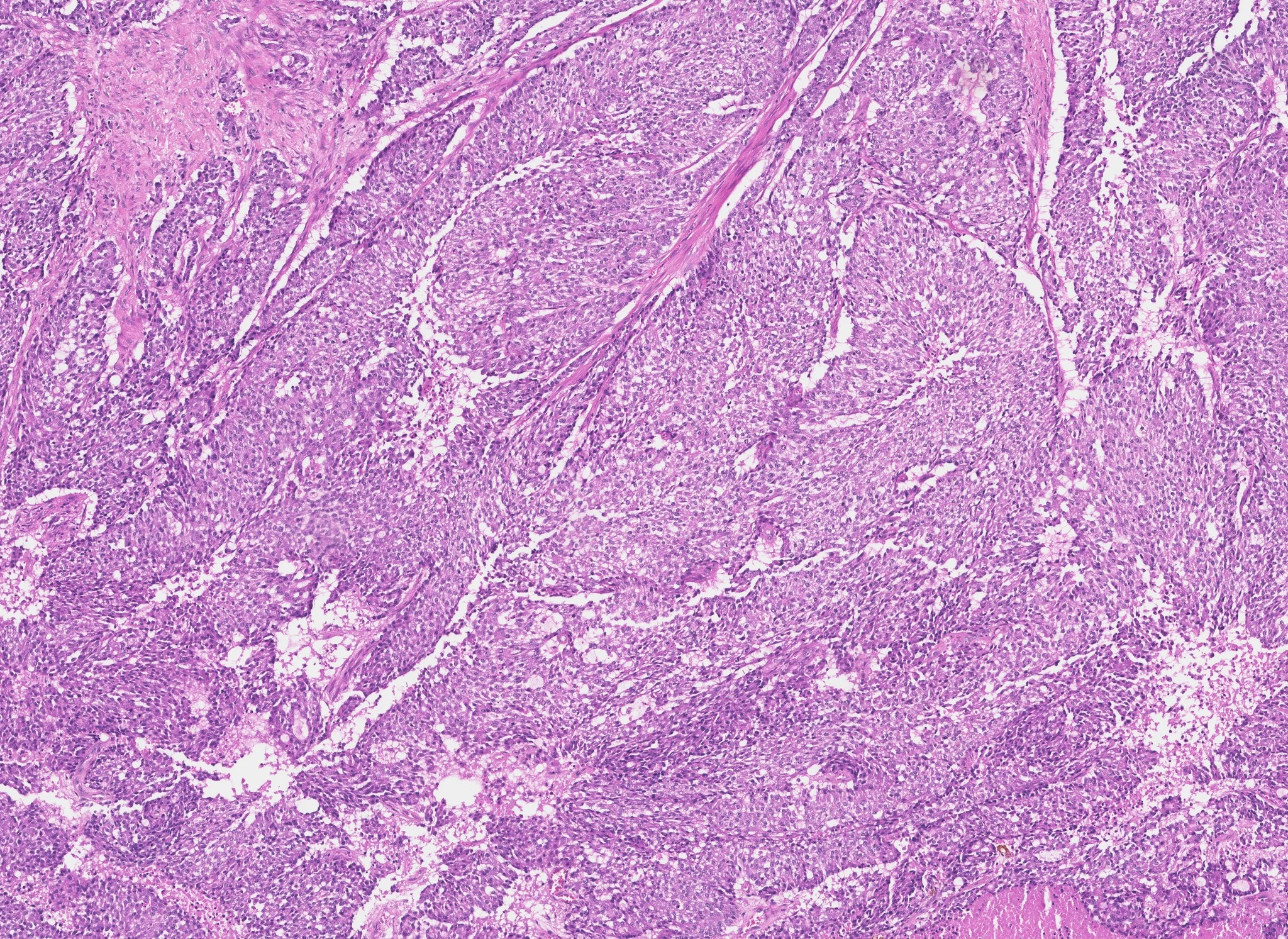
HOW IT WORKS
From slide
to reviewable score.
Whole-slide image
Access the slide for quantification directly through your supported image management system.
Mitosis
Detection, classification, heatmaps and hotspot-based mitotic scores.
Reviewable quantification result
The assay returns the score and supporting counts alongside the heatmap for expert review.
AVAILABLE ON




PUBLICATIONS AND ABSTRACTS
Evidence for Mitosis.
Studies and congress abstracts listing Mitosis as a biomarker. The full list is on the Resources page.
All publications ↗︎FAQ
Practical
questions.
01What does DiaKwant Mitosis report?
DiaKwant Mitosis reports a mitotic count from H&E-stained breast carcinoma sections.
02Which tissue and staining are covered?
The assay covers H&E-stained breast carcinoma sections.
03How is the hotspot defined?
The hotspot is the 2 mm² region of highest mitotic activity identified on the slide, and the count is performed within that region.
04What information can the pathologist review?
The pathologist can review the detected mitotic figures and the hotspot region behind the reported count.
05Does DiaKwant replace the pathologist?
No. DiaKwant is a diagnostic aid. The result must be reviewed by a qualified pathologist, who retains full responsibility for the final interpretation and report.
06How can a laboratory confirm compatibility?
Technical compatibility depends on your laboratory's scanner and image management configuration. Contact the DiaDeep team to confirm compatibility for your validated setup.
CLUB QUANTI
See how DiaKwant® handles the cases you encounter in your practice.
Compare its findings with your own scoring on your slides.
Try it on your cases ↗︎Apply to Club Quanti ↗︎