.png)
DiaKwant™ AI quantifies PD-L1 CPS/TPS, HER2, Ki-67 and more. Combined with your expertise, is what leads to better outcomes*.
Moving beyond subjective estimation to objective and cell-by-cell quantification

DiaKwant™ is validated across multiple tumor types, including NSCLC, SCLC, GC/GEJ, HNSCC, CC, and TNBC.
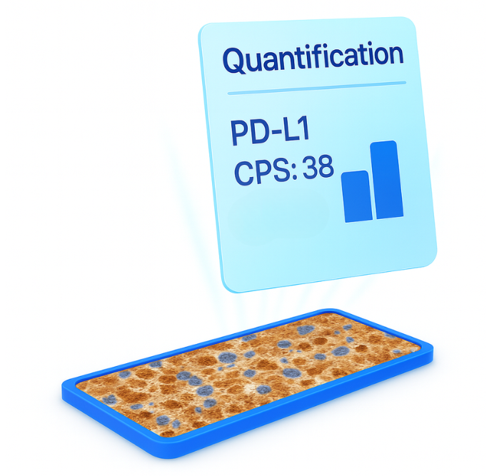
Automatically detects tumor regions and the surrounding microenvironment on IHC slides. DiaKwant™ algorithm identifies tumor and immune cells, quantifies PD-L1–positive cells, and computes CPS and TPS at the whole-slide level¹.
Scientific results showed that AI-assisted PD-L1 scoring improved accuracy (89% vs. 75% with routine methods), sensitivity (96% vs. 78%), and reproducibility across multiple carcinoma types, particularly in challenging CPS ranges¹.
DiaKwant™ detects invasive tumor regions and performs cell- and membrane-level analysis on IHC slides. The algorithm quantifies staining intensity (0, 1+, 2+, 3+) and membrane completeness across tumor cells, providing an interpretable whole-slide HER2 score in line with ASCO/CAP and GEFPICS guidelines.
In a study presented at ESMO, DiaKwant™ HER2 scoring algorithm achieved 90% balanced accuracy, with 100% detection of HER2 3+ cases and 93% of HER2-Low cases, while improving inter-observer agreement from 57% to 75%².


DiaKwant™ Ki67 automatically detects invasive tumor regions on IHC slides and quantifies proliferation at both the hotspot and whole-slide level. DiaKwant™ algorithm identifies tumor nuclei, exclude lymphocytes, classifies Ki67-positive versus total cells, and computes proliferation indices.
Results obtained showed that DiaKwant™ Ki67 model achieved high concordance with pathologist consensus, with an R² of 95% between predicted and ground truth proliferation indices. DiaKwant™ automatically detected hotspots and quantified proliferation at both hotspot and whole-slide levels³.
DiaKwant™ Mitosis automatically detects invasive tumor regions on H&E slides and identifies mitotic figures.
It highlights candidate mitoses, classifies them, and generates interpretable heatmaps and hotspot-based mitotic scores.
DiaKwant™ mitosis model reached a precision of 88% and a recall of 71%. Pathologists validated 80% of DiaKwant™ mitoses, leading to a 41% increase in consensus mitoses count and a 26% increase in individual pathologist counts⁴.
.png)

DiaKwant™ ER/PR automatically detects invasive tumor regions and performs nuclear-level analysis on IHC slides. DiaKwant™ algorithm quantifies staining intensity and percentage of positive tumor nuclei, providing interpretable whole-slide scores.
Our evaluation study showed that the DiaKwant™ ER/PR model achieved high concordance with pathologist consensus, with an R² of 0.95 between predicted and ground truth nuclear positivity. DiaKwant™ automatically detected invasive regions and quantified ER/PR expression at both hotspot and whole-slide levels³.


No manual steps needed


Identification of the regions of interest with no manual annotation


Automatic detection of cells and staining mesurement


You own your diagnostic decision, supported by objective quantification


Whether you want to test our AI on your slides and explore our solutions, we’d love to hear from you.